
3D Reconstruction of Somatogastric Ganglion Neuron
Farzan Nadim, Jorge Golowasch, Dirk Bucher

Implanted Microelectrodes Activated by Floating Lights
Mesut Sahin

Namas Chandra

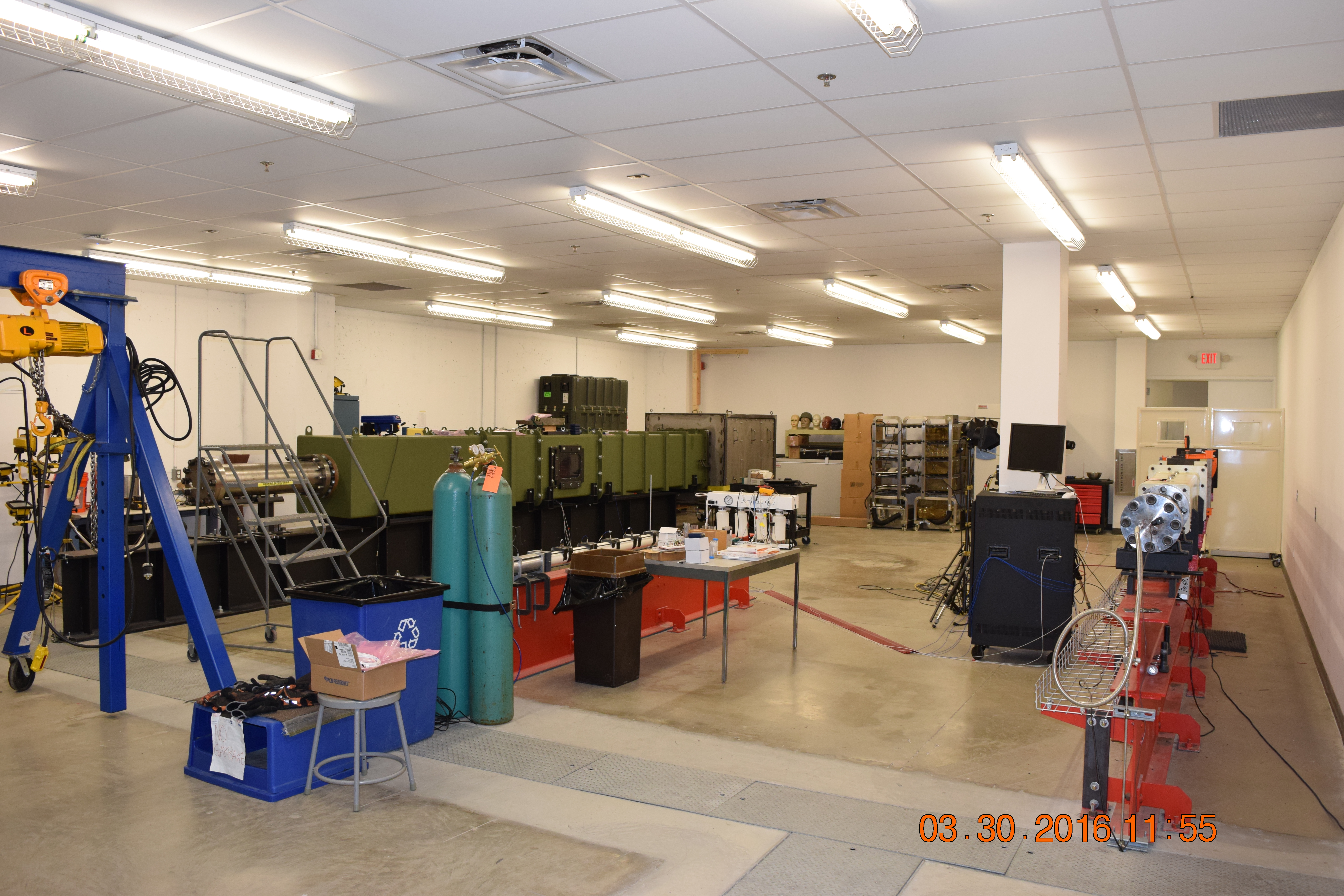
 New Generation Helmet Testing for Protection Against Blasts CIBM3
Namas Chandra
IBNR encompasses basic, applied and translational neuroscience research of faculty members from a number of NJIT departments in the Newark College of Engineering (NCE) and the College of Science and Liberal Arts (CSLA).
IBNR has two primary aims: to promote high-quality interdisciplinary research by developing synergies among research groups and to train graduate students and postdoctoral fellows in an interdisciplinary environment so that they can compete in the academic and industry markets.
The research goals of IBNR focus on fostering collaborations through seed grants and sponsoring of graduate students involved in interdisciplinary research. The IBNR runs a neuroscience-themed seminar series and sponsors a program to support visiting research scholars at NJIT, both for short durations and for sabbatical leaves.
The training program of IBNR is currently under development and involves three components. IBNR is in the process of establishing an interdisciplinary doctoral program in Neuroscience and Neural Engineering; IBNR promotes multidisciplinary undergraduate research across different Labs, Centers, and research groups; IBNR will sponsor a training workshop series for postdoctoral scholars.
In our vision, the research and training components of IBNR work jointly to promote excellence in research and training in basic and applied neuroscience by supporting the interests of faculty and students of NJIT.
Namas Chandra, Ph.D.
Distinguished Professor of Biomedical Engineering and Director
Farzan Nadim, Ph.D.
Professor of Neurobiology and Director
The purpose of the SpectraMax i3 is to provide rapid data collection of microplate readings, using the SoftMax Pro Software


Capabilities:
This device has the capability of detecting: luminescence, absorbance, and fluorescence. The system allows the user to read 6-384 well microplates simultaneously.

The SpectraMax i3 is specifically used in this lab for BCA assays and Elisa Assays. It allows the user to determine the total concentration of protein in a solution. These concentrations are then used to perform western blots. The SpectraMax i3 also allows the user to identify antibodies during Elisa Assays.
The SoftMax Pro Software quickly captures and analyzes results from the SpectraMax i3, through different data analysis options. The software has ready to run protocols to read the microplates, different curve fit options, and provides results that are ready to be used.

Purpose: To digitally scan mounted tissue samples from 1.25X to 63X magnification. Equipped with a 200-slide autoloader and a comprehensive range of image analysis solutions, the slide scanner will be used to autonomously scan holistic brain tissue, saving hours of user viewing and analysis time.
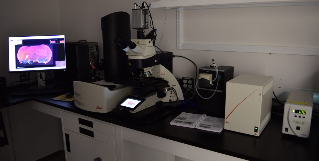
Capabilities: This microscope system is capable of viewing samples at 1.25X, 5X, 40X, and 63X magnification for both brightfield and fluorescence imaging.


Coronal brain slice of injured rat model. Blue stain represents cell nuclei (DAPI), green stain represents neurons (NeuN), and red stain represents area of extracellular matrix degradation (MMP2).
Unique Features
- 200 slide autoloader
- 1.5X, 5X, 40X, 63X magnification
- Automated oiler and oil-immersion lenses
- One-click scan protocols
- 7 filter positions
Working in uniquely developed facilities, the Institute's team of researchers at NJIT examines and solves the complex questions of brain injury from protection, diagnostics, therapeutics and clinical management perspectives.


Current lower extremity exoskeletons are both under-actuated and lack effective user control that allows independent ambulation. Rather than employ computer-generated patterns, the movements of the hands will be used as analogs of user-generated foot patterns. With the hands rigidly linked to the feet, the forces generated by the hands will directly operate an admittance controlled exoskeleton as well as receive feedback.


A 1/2 scale robot controlled by a pair of trekking poles is being evaluated in the laboratory. A human-scale admittance-controlled exoskeleton is being developed with six degrees of freedom in each leg, with force-measuring trekking poles and sensor-based ground force detection.
Combining exoskeleton therapy with simultaneous spinal cord stimulation to enhance neurorehabilitation of individuals with incomplete spinal cord injury.


Current Projects
- Hand Rehabilitation Post-Stroke Using Interactive Virtual Environments
- Rehabilitation Engineering Research Center on Wearable Robots
Visit the Center for Rehabilitation Robotics
Our laboratory is developing a floating light activated micro electric stimulator (FLAMES) that is a wireless implantable device for neural stimulation where near-infrared (NIR) light is used for energy transfer to the microstimulator through neural tissue.
The FLAMES was acutely tested in the rat spinal cord for feasibility of the main concept. Temperature elevation profile was also measured experimentally using a micro termoprobe inside the rat brain induced by an NIR laser beam to determine the maximum allowable optical power. Our latest chronic implants have shown minimal tissue response to untethered devices implanted into the brain and the spinal cord in rats.
These studies so far produced supportive evidence for a wireless microstimulator that can be activated optically by NIR light through neural tissue. Devices at submillimeter scale were designed for wireless activation of neurons in the central nervous system. Prototypes were fabricated in collaboration with Dr. Unlu's group at Boston University.